What Are The Different Parts of Your Eye?
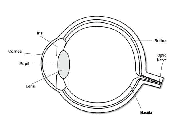
The human eye is made up of many different parts that work together to create the sense of sight. The major components include the cornea, lens, iris, pupil, retina, and optic nerve.
Cornea:
The cornea is the transparent outer layer of the eye that covers the front. Its job is to help focus light as it enters the eye. The cornea is curved, allowing it to bend (refract) light, guiding it toward the lens.
Lens:
Located just behind the pupil, the lens is responsible for fine-tuning focus. It changes shape depending on whether the object you’re looking at is near or far. The lens is flexible and works with the ciliary muscles to adjust its thickness, which allows for clear vision at different distances.
Iris:
The iris is the colored part of the eye surrounding the pupil. Its primary function is to control the amount of light that enters the eye by expanding or contracting the pupil. In bright light, the iris contracts the pupil to reduce the amount of light entering, while in low light, it widens to allow more light in.
Pupil:
The pupil is the black circular opening in the center of the iris. It controls how much light reaches the retina. In low light conditions, the pupil enlarges to let in more light; in bright light, it constricts.
Retina:
The retina is a light-sensitive layer at the back of the eye. It contains photoreceptor cells called rods and cones. Rods help you see in dim light and detect movement, while cones are responsible for color vision and sharp detail. The retina converts light into electrical signals, which are then transmitted to the brain through the optic nerve.
Macula:
The macula is the central part of the retina responsible for sharp, central vision. It allows you to see fine details, such as when reading or recognizing faces.
Optic Nerve:
The optic nerve carries the electrical signals from the retina to the brain. The brain then interprets these signals as images, giving you the ability to see.
How The Human Eye Works
When light enters the eye through the cornea, the cornea bends or refracts the light, directing it through the pupil. The amount of light that enters is regulated by the iris, which expands or contracts to adjust the size of the pupil, depending on the brightness of the surroundings.
Once the light has passed through the pupil, it reaches the lens. The lens changes shape, flattening or thickening, to focus the light onto the retina. This process, called accommodation, is what allows you to see both near and distant objects clearly.
The retina is where the actual process of turning light into visual information begins. The retina contains millions of cells, including two types of photoreceptor cells: rods and cones. Rods are responsible for vision in low light conditions and help with seeing shapes and movement. Cones are concentrated in the center of the retina, called the macula, and are responsible for color vision and seeing fine details.
When light hits these photoreceptors, it is converted into electrical signals. The retina processes these signals and sends them through the optic nerve to the brain. This process happens almost instantaneously, allowing us to see in real-time.
What Are Common Vision Problems?
Sometimes the eye doesn’t function perfectly, leading to vision problems. These conditions usually occur due to the way light is refracted within the eye.
Nearsightedness (Myopia):
Myopia occurs when the eyeball is too long or the cornea is too curved. This causes light to focus in front of the retina, making distant objects appear blurry. People with myopia often have difficulty seeing things far away but can see close objects clearly.
Farsightedness (Hyperopia):
Hyperopia is the opposite of myopia. It happens when the eyeball is too short or the cornea is too flat, causing light to focus behind the retina. This makes close objects appear blurry, while distant objects are usually seen more clearly.
Astigmatism:
Astigmatism occurs when the cornea or lens is irregularly shaped. Instead of being round like a basketball, the surface is more oval, like a football. This irregular shape causes light to scatter, leading to distorted or blurred vision. Astigmatism can affect both near and far vision.
Presbyopia:
Presbyopia is an age-related condition where the lens loses flexibility. As a result, it becomes harder to focus on close objects. This typically begins in people over the age of 40, and many need reading glasses or bifocals to see things up close.
Cataracts:
A cataract is the clouding of the eye’s natural lens, often due to aging. This cloudiness blocks or scatters light, causing blurry vision, glare, and difficulty seeing at night. Cataracts can be treated with surgery to remove the clouded lens and replace it with an artificial one.
Macular Degeneration:
This condition affects the macula, the central part of the retina responsible for sharp vision. Macular degeneration leads to a gradual loss of central vision, making tasks like reading and recognizing faces difficult. It’s more common in older adults and is a leading cause of vision loss.
Glaucoma:
Glaucoma is a group of eye conditions that damage the optic nerve, often due to high pressure inside the eye. If left untreated, glaucoma can lead to vision loss and blindness. Regular eye exams are important for detecting and managing glaucoma early.
Photokeratitis:
Often described as “sunburn of the eyes,” photokeratitis is a temporary but painful condition caused by overexposure to UV rays. Symptoms include redness, pain, and a gritty feeling in the eyes. It typically heals within a few days but can be prevented by wearing UV-protective sunglasses.
Discover independent eyewear with Project Spex. Every Friday, we deliver the latest in collectible eyewear and your favorite independent designers.
Sign up now and never miss a thing!
About The Author:
Will Benjamin is an advocate for independent eyewear and one of the driving forces behind Project Spex. With a passion for unique, collectible, and limited-edition eyewear, Will aims to inspire people to build their own collections through Project Spex, while supporting the success of independent opticals.